
Breast Cancer Model Manufacturer,Supplier and Exporter
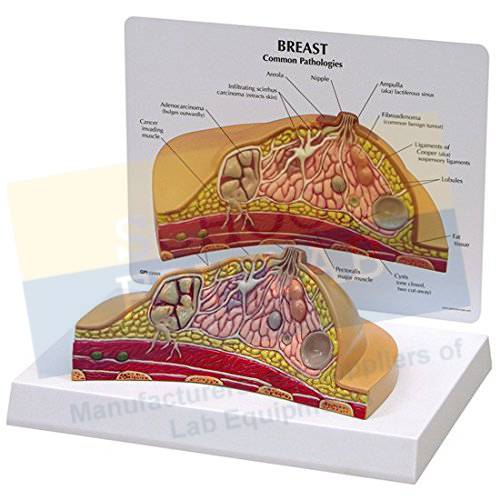
Product Code : SEL-AM-10105
Quick Answer: Shown as a full-size cross-section on a base, the Breast Cancer Model depicts adenocarcinoma, cysts, fibroadenoma and infiltrating scirrhus carcinoma together with breast structures such as suspensory ligaments, fat tissue, lymph nodes, muscles and ribs.
Last updated: October 2026
Key Features
- Full-size cross-section model
- Depicts adenocarcinoma, cysts, fibroadenoma and infiltrating scirrhus carcinoma
- Shows suspensory ligaments, fat tissue, lymph nodes, muscles and ribs
- Mounted on a base
- Size 16 x 13 x 12 cm
Technical Specifications
| Specification | Value |
|---|---|
| Type | Full-size cross-section |
| Pathologies depicted | Adenocarcinoma, cysts, fibroadenoma, infiltrating scirrhus carcinoma |
| Structures shown | Suspensory ligaments, fat tissue, lymph nodes, muscles, ribs |
| Mounting | Base |
| Size | 16 x 13 x 12 cm |
About This Product
In anatomy and pathology teaching, a sectioned model lets students compare the appearance of several breast pathologies on one piece. The cross-section places each condition among the suspensory ligaments, fat tissue, lymph nodes, muscles and ribs, so the class can relate it to the structures around it. The Breast Cancer Model can be packed for export to Timor-Leste, Suriname, Chile, the Bahamas, Mexico and Italy.
Frequently Asked Questions
Which pathologies does the breast cancer model depict?
It depicts common pathologies including adenocarcinoma, cysts, fibroadenoma and infiltrating scirrhus carcinoma in a full-size cross-section.
Which breast structures are shown?
The model shows suspensory ligaments, fat tissue, lymph nodes, muscles and ribs. It is mounted on a base and measures 16 x 13 x 12 cm.
Related: Anatomical Models, Colon Model With Pathologies, Diabetic Foot Model.
For a quotation on the Breast Cancer Model, contact School Educational Instrument.


