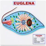
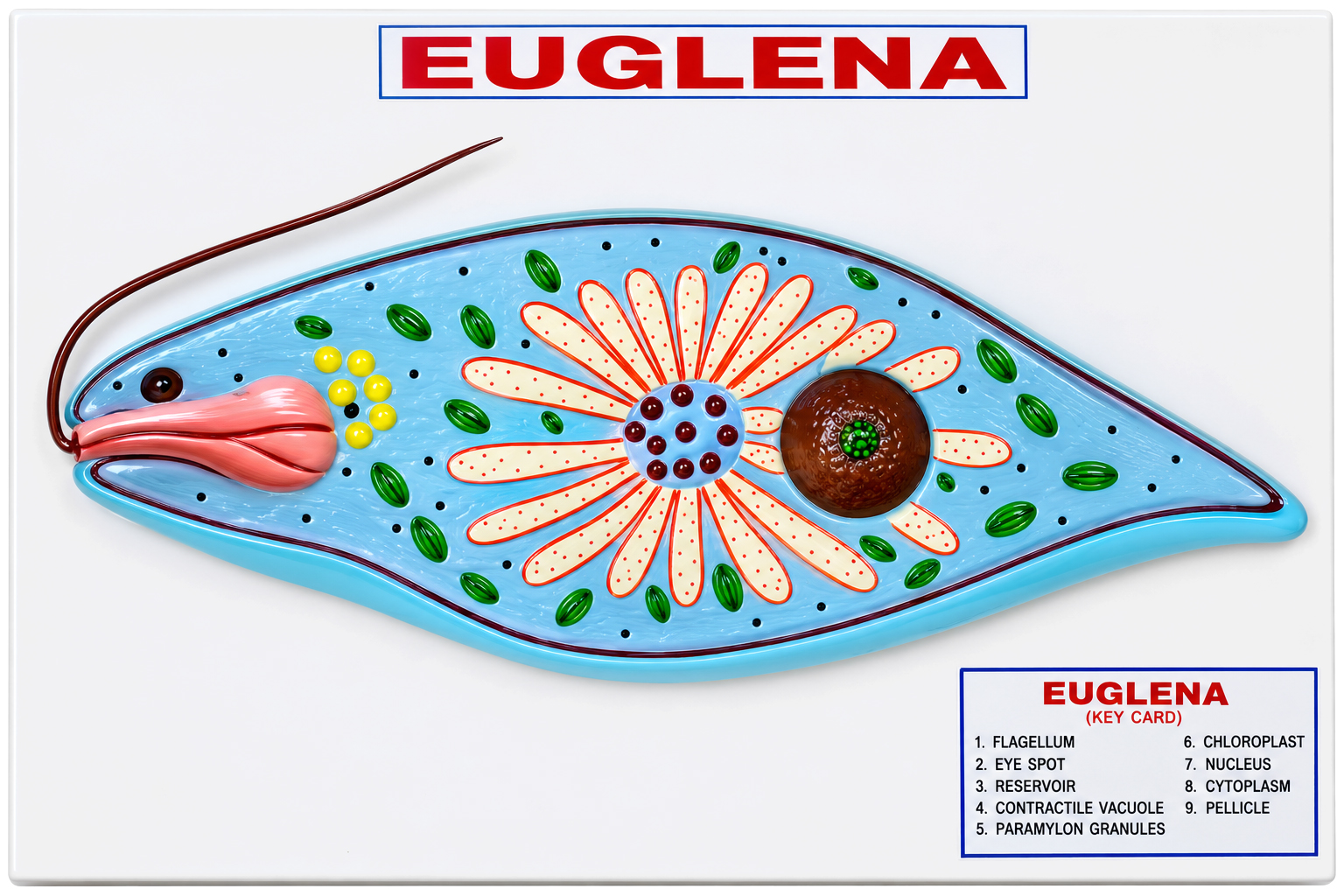
Quick Answer: For lessons on single-celled organisms, the Euglena Model gives an enlarged view of the cell with its flagellum, stigma, chloroplast, paramylum bodies and contractile vacuole numbered against an English key card.
Last updated: October 2026
Key Features
Enlarged view of a Euglena cell
Flagellum, basal granule, reservoir and cytostome
Stigma, chloroplast and paramylum bodies
Nucleus with endosome, contractile vacuole and pellicle
Mounted on a base, numbered with an English key card
About This Product
Euglena is a familiar example for showing that a single cell can combine plant-like and animal-like features. On this model the chloroplast and paramylum bodies appear alongside the flagellum, reservoir and contractile vacuole, so a teacher can talk through feeding, movement and the cell surface from one display. Each part carries a number that matches the English key card, useful for labelling practice in school and college biology.
Frequently Asked Questions
Which parts of Euglena are identified?
The cytostome, stigma, reservoir, flagellum, basal granule, paramylum bodies, chloroplast, contractile vacuole, nucleus, endosome and pellicle are all numbered and listed on the English key card.

 Model.jpg)