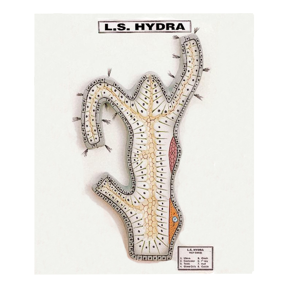
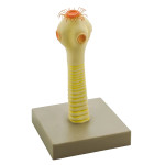
Quick Answer: Colourfully illustrated and mounted on a stand, the Hydra Model shows the external and internal structure of a hydra, from the tentacles, hypostome and bud to the mesoglea, nematocysts and flagellated cell, numbered with an English key card.
Last updated: October 2026
Key Features
External and internal structure of a hydra
Bud, tentacle, hypostome, mouth and foot
Gastrovascular cavity, mesoglea, epidermis and gastrodermis
The body layers and cell types of hydra are hard to make out on a slide, so an illustrated model gives students a clear reference. They can find the epidermis, mesoglea and gastrodermis, then pick out the nematocysts, bud, testis and ovary, checking each against the numbered English key card. Buyers in Mali, Croatia, Gabon, Samoa, South Sudan and France can send an export enquiry for the Hydra Model.
Frequently Asked Questions
Which structures are shown on the hydra model?
The model shows the bud, gastrovascular cavity, testis, tentacle, foot, ovary, hypostome, mesoglea, mouth, nematocysts, epidermis, gastrodermis and flagellated cell, numbered with an English key card.